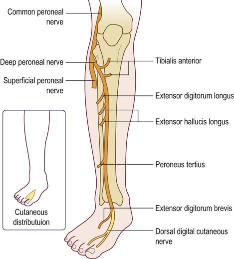
There are two cutaneous branches that arise directly from the common fibular nerve as it moves over the lateral head of the gastrocnemius. If the common fibular nerve is damaged, the patient may lose the ability to dorsiflex, evert the foot, and extend the digits.

It also innervates some intrinsic muscles of the foot. These muscles act to dorsiflex the foot, and extend the digits. Deep fibular nerve: Innervates the muscles of the anterior compartment of the leg tibialis anterior, Extensor Digitorum Longus and extensor hallucis longus.Superficial fibular nerve: Innervates the muscles of the lateral compartment of the leg fibularis longus and brevis.In addition, its terminal branches also provide innervation to muscles: The common fibular nerve innervates the short head of the biceps femoris muscle (part of the hamstring muscles, which flex at the knee).
#Palpation n peroneus skin#
Also supplies (via branches) cutaneous innervation to the skin of the anterolateral leg, and the dorsum of the foot. Sensory: Innervates the skin over the upper lateral and lower posterolateral leg. Also supplies (via branches) the muscles in the lateral and anterior compartments of the leg. Motor: Innervates the short head of the biceps femoris directly.

It usually arises in common with the fibular (peroneal) communicating branch which joins the sural nerve in the middle third of the leg. The lateral cutaneous nerve of the calf supplies the posterolateral side of the proximal two-thirds of the leg.

The common fibular (peroneal) nerve gives articular branches to the knee and superior tibiofibular joint. The nerve can be palpated behind the head of the fibula and as it winds around the neck of the fibula. It passes forwards around the neck of the fibula within the substance of fibularis (peroneus) longus, where it terminates by dividing into the superficial and deep fibular (peroneal) nerves. It courses along the upper lateral side of the popliteal fossa, deep to biceps femoris and its tendon until it gets to the posterior part of the head of the fibula. The common peroneal nerve is the smaller and terminal branch of the sciatic nerve which is composed of the posterior divisions of L4, 5, S1, 2.


 0 kommentar(er)
